›Questions
OphthalmologyVisual Field Testing
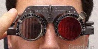
Q465. Following test is used to diagnose which ocular condition as shown in image:
Expand
Tap an option to reveal the answer
Q465. Following test is used to diagnose which ocular condition as shown in image:
Tap an option to reveal the answer
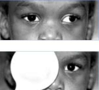
Q466. Name the manoeuvre shown in the image:

Tap an option to reveal the answer
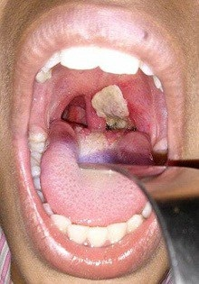
Q467. Identify the condition of the given image:
Tap an option to reveal the answer
Q468. CT image of left maxilla with history of allergic rhinitis?
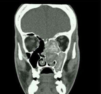
Tap an option to reveal the answer
Q469. Identify the instrument:
Tap an option to reveal the answer
Q470. Identify the lesion of vocal cord in the image given below:
Tap an option to reveal the answer
Q471. A patient gives H/o hoarseness in voice & presenting with clinical condition as shown in the image. Identify the lesion:
Tap an option to reveal the answer
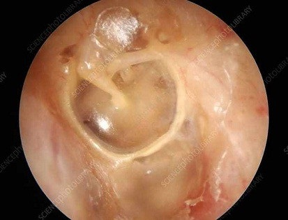
Q472. A patient presented with the following picture of Tympanic Membrane. Most Probable diagnosis (marked with arrow):
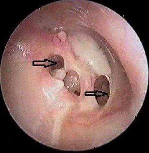
Tap an option to reveal the answer
Q473. Which method is used to study the following timeline?

Tap an option to reveal the answer
Q474. A 6 year old boy is having symptoms such as fever and chills, cough, rapid breathing, difficulty breathing, and chest pain, culture from a sample shows Gram-positive culture, Identify the image:
Tap an option to reveal the answer
Q439. Identify the infection from the chest X- ray of a patient with low-grade fever?
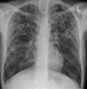
Tap an option to reveal the answer
Q475. 12 Identify the picture

Tap an option to reveal the answer
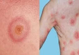
Q476. Identify the image
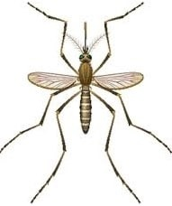
Tap an option to reveal the answer
Q477. What is the cause of death according to the below death certificate
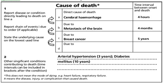
Tap an option to reveal the answer
Q478. Identify the blood grouping process done here. Slide given with: AB no clumps, Clumping in O & No clumps in control. Which group does this signify?
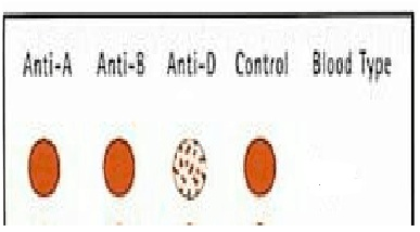
Tap an option to reveal the answer
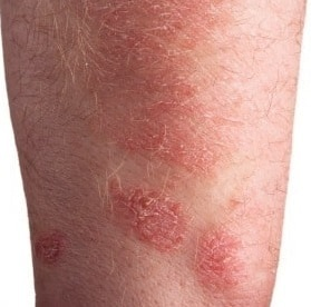
Q479. Identify the condition as shown in the image given below:
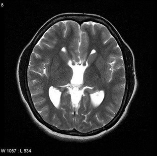
Tap an option to reveal the answer
Q480. Which nerve supplies to the area marked as ‘Area B’ in the image:
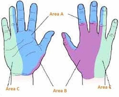
Tap an option to reveal the answer
Q481. Which of the following is not a boundary of given image:
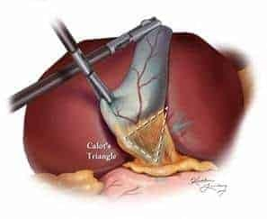
Tap an option to reveal the answer
Q482. Marked area in the given image is supplied by which dermatome?
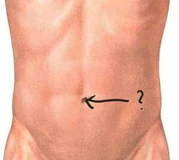
Tap an option to reveal the answer
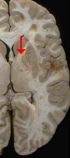
Q483. Identify the type of the fibre marked in the image of internal capsule:
Tap an option to reveal the answer