NEET-PG PYQ logoNEET-PG PYQ
SubjectsTopicsYearsGo Premium
© 2026 NEET PG PYQ. All rights reserved.
Home
›Subjects
›Year 2019
›Radiology
RadiologyMusculoskeletal Radiology

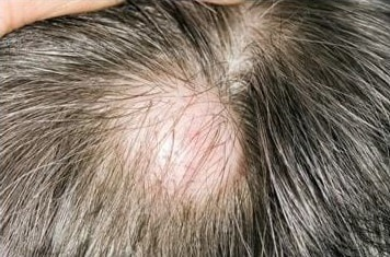
Q447. Identify the condition shown in the image?

Clinical photograph of the scalp showing patchy, well-defined, non-scarring hair loss consistent with alopecia areata
Expand

Tap an option to reveal the answer

NEET-PG2019Repeats: N/A
Radiology
Musculoskeletal Radiology

Q451. Identify the condition as shown:
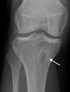
X-ray of the knee joint showing a lytic lesion in the proximal tibia with an arrow pointing to a well-defined cavity consistent with Brodie abscess
Expand

Tap an option to reveal the answer

NEET-PG2019Repeats: N/A
RadiologyUrological Radiology

Q456. A female patient presented with recurrent Urinary tract infections. Imaging shows the following picture. What can be the most probable diagnosis?
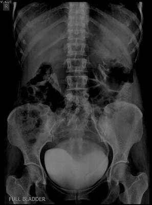
IVP/X-ray of the abdomen showing a cobra head deformity of the ureter at the vesico-ureteric junction consistent with ureterocele
Expand

Tap an option to reveal the answer

NEET-PG2019Repeats: N/A
RadiologyChest Radiology

Q439. Identify the infection from the chest X- ray of a patient with low-grade fever?
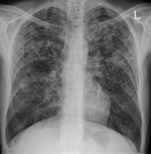
Chest X-ray (PA view) showing bilateral diffuse miliary nodular opacities throughout both lung fields consistent with miliary tuberculosis
Expand

Tap an option to reveal the answer

NEET-PG2019Repeats: N/A
RadiologyNeuroradiology

Q479. Identify the condition as shown in the image given below:
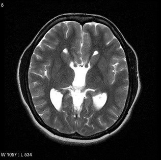
Axial MRI of the brain showing widely spaced lateral ventricles with a 'racing car sign' consistent with callosal dysgenesis/agenesis of corpus callosum
Expand

Tap an option to reveal the answer

NEET-PG2019Repeats: N/A
RadiologyGastrointestinal Radiology

Q442. Identify the radiological procedure shown below?
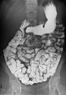
X-ray image of the abdomen showing contrast outlining the entire small bowel in a barium meal follow-through study
Expand

Tap an option to reveal the answer

NEET-PG2019Repeats: N/A
RadiologyGastrointestinal Radiology

Q443. Identify the condition in the below image?
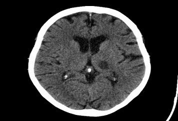
Axial CT scan of the brain showing a small hyperdense lesion in the thalamic/basal ganglia region consistent with a lacunar infarct
Expand

Tap an option to reveal the answer

NEET-PG2019Repeats: N/A
RadiologyContrast Media

Q430. Which of the following is water soluble contrast?

Tap an option to reveal the answer

NEET-PG2019Repeats: N/A
RadiologyBone Radiology

Q431. Solitary lytic lesions seen in

Tap an option to reveal the answer
NEET-PG2019Repeats: N/A
Showing 1-9 of 9 questions
for subject "Radiology"

from year 2019