NEET-PG PYQ logoNEET-PG PYQ
SubjectsTopicsYearsGo Premium
© 2026 NEET PG PYQ. All rights reserved.
Home
›Subjects
›Year 2019
AnatomyAbdominal Anatomy

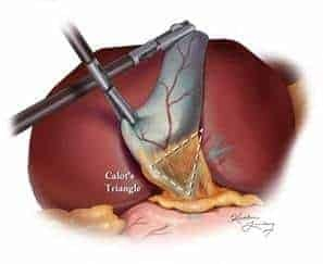
Q481. Which of the following is not a boundary of given image:

Intraoperative photograph showing the Calot's triangle (cystohepatic triangle) during cholecystectomy, bounded by the common hepatic duct, cystic duct, and inferior surface of liver
Expand

Tap an option to reveal the answer

NEET-PG2019Repeats: N/A
AnatomyUpper Limb Anatomy

Q482. Marked area in the given image is supplied by which dermatome?
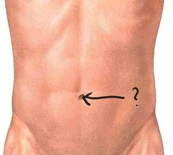
Anatomical diagram of the anterior abdominal wall with an arrow pointing to the umbilical region, marking the T10 dermatome level
Expand

Tap an option to reveal the answer

NEET-PG2019Repeats: N/A
AnatomyNeuroanatomy

Q483. Identify the type of the fibre marked in the image of internal capsule:
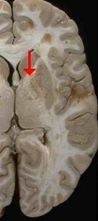
Gross specimen of the brain showing a coronal section with an arrow (red) pointing to the internal capsule between the thalamus and lentiform nucleus
Expand

Tap an option to reveal the answer

NEET-PG2019Repeats: N/A
AnatomyNeuroanatomy

Q484. Identify the structure marked by a red arrow in the given image:
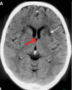
Axial CT scan of the brain showing a midline structure with a red arrow indicating the fornix (C-shaped white matter tract connecting hippocampus to mammillary bodies)
Expand

Tap an option to reveal the answer

NEET-PG2019Repeats: N/A
MedicineCardiology

Q440. Identify the condition given in the image?
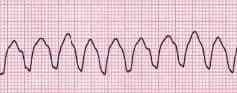
ECG strip showing regular wide-complex tachycardia with uniform QRS morphology consistent with ventricular tachycardia
Expand

Tap an option to reveal the answer

NEET-PG2019Repeats: N/A
AnatomyUpper Limb Anatomy

Q485. What is the Nerve Supply of marked structure in the given image:
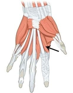
Anatomical diagram of the palmar surface of the hand showing an arrow pointing to the first lumbrical muscle at the lateral aspect of the index finger
Expand

Tap an option to reveal the answer

NEET-PG2019Repeats: N/A
AnatomyNeuroanatomy

Q486. Identify the marked structure in the image:
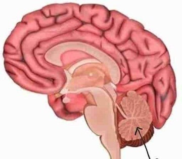
Anatomical diagram of a sagittal section of the brain with a black arrow pointing to the cerebellum at the posterior fossa
Expand

Tap an option to reveal the answer

NEET-PG2019Repeats: N/A
ENTParotid Region

Q487. Nerve injured in Frey’s syndrome is:
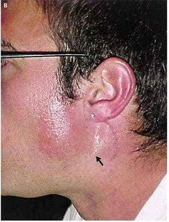
Clinical photograph of the face showing flushing/erythema of the pre-auricular region and cheek on one side consistent with Frey's syndrome (auriculotemporal nerve injury)
Expand

Tap an option to reveal the answer

NEET-PG2019Repeats: N/A
AnatomyHead and Neck Anatomy

Q488. Which nerve passes through the marked foramen in the given image:
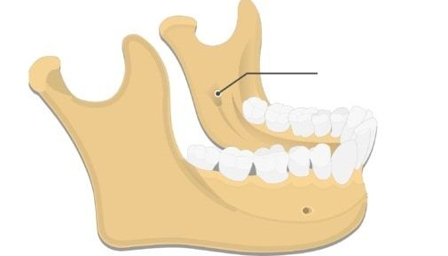
Anatomical diagram of the mandible showing the mandibular foramen on the medial surface of the ramus with a line pointer indicating it
Expand

Tap an option to reveal the answer

NEET-PG2019Repeats: N/A
AnatomyUpper Limb Anatomy

Q489. Identify the marked muscle ‘A’ in the diagram:
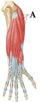
Anatomical diagram of the posterior forearm/dorsum of hand showing the marked muscle 'A' at the lateral epicondyle region consistent with extensor carpi radialis longus
Expand

Tap an option to reveal the answer

NEET-PG2019Repeats: N/A
DermatologyNail Disorders

Q490. Which of the following nail findings is seen in the condition shown below?
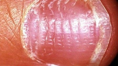
Clinical photograph of a fingernail showing longitudinal ridging/pitting on the nail surface consistent with nail pitting (seen in psoriasis/alopecia areata)
Expand

Tap an option to reveal the answer

NEET-PG2019Repeats: N/A
OphthalmologyOrbit

Q441. A patient presented with history of diplopia and restricted eye movements. A clinical image and CT image is shown below. What will be the diagnosis?
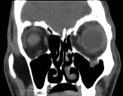
Coronal CT scan of the face showing blowout fracture of the orbital floor/wall with herniation of orbital contents into the maxillary sinus
Expand

Tap an option to reveal the answer

NEET-PG2019Repeats: N/A
RadiologyGastrointestinal Radiology

Q442. Identify the radiological procedure shown below?
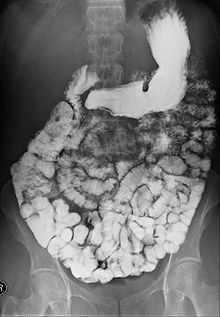
X-ray image of the abdomen showing contrast outlining the entire small bowel in a barium meal follow-through study
Expand

Tap an option to reveal the answer

NEET-PG2019Repeats: N/A
RadiologyGastrointestinal Radiology

Q443. Identify the condition in the below image?
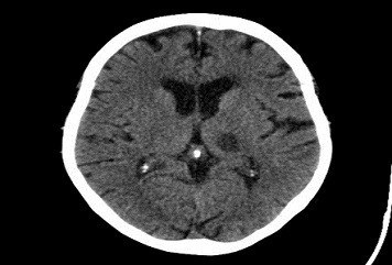
Axial CT scan of the brain showing a small hyperdense lesion in the thalamic/basal ganglia region consistent with a lacunar infarct
Expand

Tap an option to reveal the answer

NEET-PG2019Repeats: N/A
DermatologyPhotodermatology

Q444. A child with history of fever, photosensitivity, rash sparing nasolabial fold presents to OP. Identify the condition?
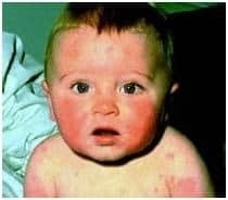
Clinical photograph of an infant with erythematous malar rash sparing the nasolabial fold consistent with neonatal SLE
Expand

Tap an option to reveal the answer

NEET-PG2019Repeats: N/A
MedicineNeurology

Q198. Syndrome associated with posterior inferior cerebellar artery thrombosis:

Tap an option to reveal the answer

NEET-PG2019Repeats: N/A
AnatomyAbdominal Anatomy

Q199. Space of Disse is in:

Tap an option to reveal the answer

NEET-PG2019Repeats: N/A
PathologyNeuropathology

Q200. What are Gitter cells:

Tap an option to reveal the answer

NEET-PG2019Repeats: N/A
AnatomyAbdominal Anatomy

Q201. Cholecystocaval line separates:

Tap an option to reveal the answer

NEET-PG2019Repeats: N/A
PathologyGeneral Pathology

Q202. Which of the following exocrine glandular ducts are not obstructed in cystic fibrosis:

Tap an option to reveal the answer
NEET-PG2019Repeats: N/A
PreviousNext
Previous
12345
Next
Showing 41-60 of 320 questions
from year 2019