NEET-PG PYQ logoNEET-PG PYQ
SubjectsTopicsYearsGo Premium
© 2026 NEET PG PYQ. All rights reserved.
Home
›Subjects
›Year 2019
SurgeryVascular Surgery

Q463. A patient came in emergency with severe abdominal pain pulse 112/ minute and systolic BP 80 mm/Hg with the chest X- ray shown below. What will be the next appropriate step to be taken for the patient?
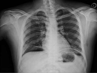
Chest X-ray showing air under the left hemidiaphragm (pneumoperitoneum) consistent with hollow viscus perforation
Expand

Tap an option to reveal the answer

NEET-PG2019Repeats: N/A
Ophthalmology
Retina

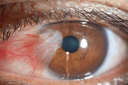
Q464. True statement about given condition of eye except:

Clinical photograph of the eye showing a wing-shaped fibrovascular growth from the conjunctiva encroaching onto the cornea consistent with pterygium
Expand

Tap an option to reveal the answer

NEET-PG2019Repeats: N/A
DermatologyPsoriasis

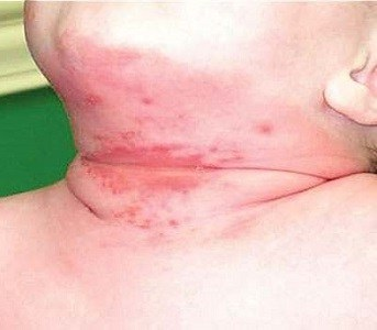
Q438. Identify the condition shown in the image below.

Clinical photograph showing an erythematous rash on the neck/chin of an infant consistent with intertrigo
Expand

Tap an option to reveal the answer

NEET-PG2019Repeats: N/A
OphthalmologyVisual Field Testing

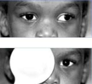
Q465. Following test is used to diagnose which ocular condition as shown in image:

Two clinical photographs demonstrating the cover-uncover test in a child with strabismus showing eye movement on covering one eye
Expand

Tap an option to reveal the answer

NEET-PG2019Repeats: N/A
ENTVertigo

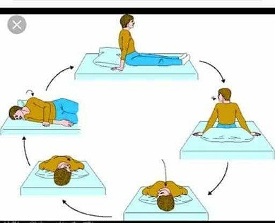
Q466. Name the manoeuvre shown in the image:

Diagram/illustration showing the Epley maneuver - a series of head positions used to treat BPPV involving rotation through multiple positions
Expand

Tap an option to reveal the answer

NEET-PG2019Repeats: N/A
ENTEar

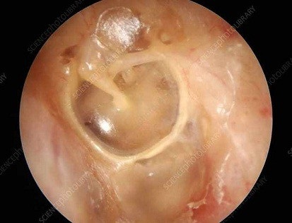
Q467. Identify the condition of the given image:

Otoscopic view showing deep widening of the ear canal with accumulation of desquamated skin (keratin plug) exposing the circular annulus consistent with keratosis obturans
Expand

Tap an option to reveal the answer

NEET-PG2019Repeats: N/A
ENTRhinology

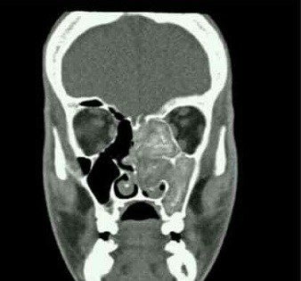
Q468. CT image of left maxilla with history of allergic rhinitis?

Coronal CT scan of the sinuses showing opacification of the left maxillary sinus with central hyperdensity (double density sign) consistent with allergic fungal sinusitis
Expand

Tap an option to reveal the answer

NEET-PG2019Repeats: N/A
SurgerySurgical Instruments

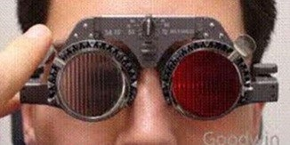
Q469. Identify the instrument:

Clinical photograph of a patient wearing the Maddox rod instrument - red cylindrical lens device used to assess and measure strabismus
Expand

Tap an option to reveal the answer

NEET-PG2019Repeats: N/A
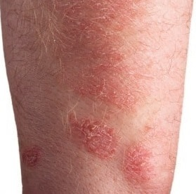
ENTLarynx

Q470. Identify the lesion of vocal cord in the image given below:

Clinical photograph showing papillomatous pink growths on the skin surface (representing laryngeal papilloma changes) with HPV-related lesions
Expand

Tap an option to reveal the answer

NEET-PG2019Repeats: N/A
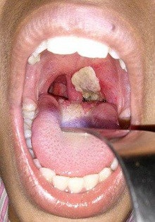
ENTLarynx

Q471. A patient gives H/o hoarseness in voice & presenting with clinical condition as shown in the image. Identify the lesion:

Clinical photograph of the oral cavity showing a thick white pseudo-membrane over the tonsil and pharynx consistent with diphtheria
Expand

Tap an option to reveal the answer

NEET-PG2019Repeats: N/A
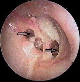
ENTEar

Q472. A patient presented with the following picture of Tympanic Membrane. Most Probable diagnosis (marked with arrow):

Otoscopic view showing multiple perforations of the tympanic membrane consistent with tubercular otitis media
Expand

Tap an option to reveal the answer

NEET-PG2019Repeats: N/A
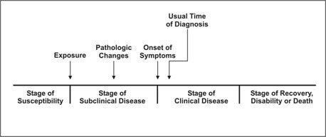
Community MedicineEpidemiology

Q473. Which method is used to study the following timeline?

Diagram showing the natural history of disease timeline with stages: susceptibility, subclinical disease, clinical disease, and recovery/disability/death
Expand

Tap an option to reveal the answer

NEET-PG2019Repeats: N/A
PaediatricsRespiratory Infections

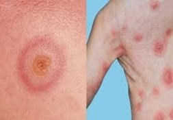
Q474. A 6 year old boy is having symptoms such as fever and chills, cough, rapid breathing, difficulty breathing, and chest pain, culture from a sample shows Gram-positive culture, Identify the image:

Two clinical photographs showing erythematous macular rash and positive alpha-hemolysis on culture plate consistent with Streptococcus pneumoniae infection
Expand

Tap an option to reveal the answer

NEET-PG2019Repeats: N/A
RadiologyChest Radiology

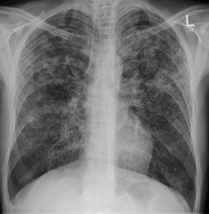
Q439. Identify the infection from the chest X- ray of a patient with low-grade fever?

Chest X-ray (PA view) showing bilateral diffuse miliary nodular opacities throughout both lung fields consistent with miliary tuberculosis
Expand

Tap an option to reveal the answer

NEET-PG2019Repeats: N/A
MicrobiologyParasitology

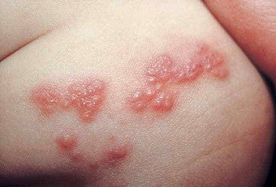
Q475. 12 Identify the picture

Clinical photograph showing grouped vesicular lesions on erythematous base in a dermatomal distribution on the trunk consistent with herpes zoster (shingles)
Expand

Tap an option to reveal the answer

NEET-PG2019Repeats: N/A
MicrobiologyParasitology

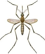
Q476. Identify the image

Illustration/diagram of the Culex mosquito (vector for Japanese encephalitis) showing characteristic long legs and striped body pattern
Expand

Tap an option to reveal the answer

NEET-PG2019Repeats: N/A
Forensic MedicineVital Statistics

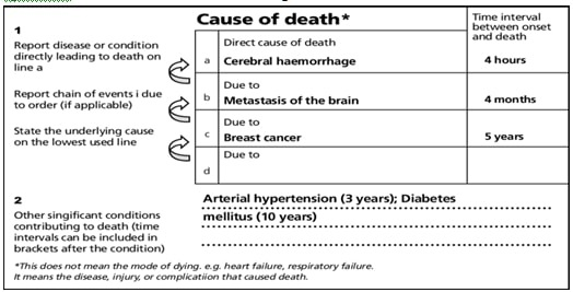
Q477. What is the cause of death according to the below death certificate

Image of a death certificate showing the cause of death sequence: Cerebral haemorrhage (4 hours) due to metastasis of the brain (4 months) due to breast cancer (5 years), with arterial hypertension and diabetes as contributory causes
Expand

Tap an option to reveal the answer

NEET-PG2019Repeats: N/A
PathologyBlood Transfusion

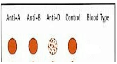
Q478. Identify the blood grouping process done here. Slide given with: AB no clumps, Clumping in O & No clumps in control. Which group does this signify?

Image of blood grouping slide test showing: Anti-A no clumps, Anti-B no clumps, Anti-D clumping, Control with oval red cells - consistent with D antigen (Rh group) testing
Expand

Tap an option to reveal the answer

NEET-PG2019Repeats: N/A
RadiologyNeuroradiology

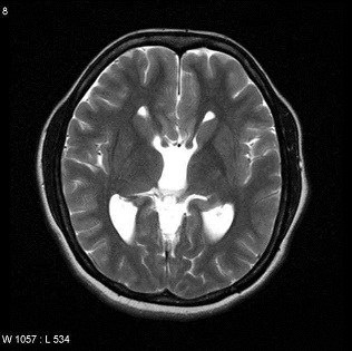
Q479. Identify the condition as shown in the image given below:

Axial MRI of the brain showing widely spaced lateral ventricles with a 'racing car sign' consistent with callosal dysgenesis/agenesis of corpus callosum
Expand

Tap an option to reveal the answer

NEET-PG2019Repeats: N/A
AnatomyUpper Limb Anatomy

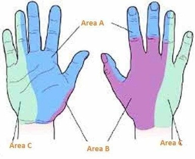
Q480. Which nerve supplies to the area marked as ‘Area B’ in the image:

Anatomical diagram of the palmar and dorsal surfaces of the hand showing color-coded nerve supply areas: Area A (ulnar nerve), Area B (radial nerve), Area C (median nerve)
Expand

Tap an option to reveal the answer

NEET-PG2019Repeats: N/A
PreviousNext
Previous
12345
Next
Showing 21-40 of 320 questions
from year 2019